|
CPT 73564 (Radiologic examination, knee;complete, four or more views. Includes professional and technical components. Requires a separate written report, signed by the interpretting physician. Interpretation must include views, anatomic location, diagnosis, reason for xray and interpretation.
CPT 73564-26 (Professional component only. Xrays must not have already been interpretted.) CPT 73564-TC (Technical component only.) |
||||||||
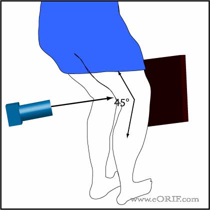 |
Weight-Bearing P/A (Rosenberg)
Position: Standing with knees flexed 45° with grid in front of knees Beam directed 10° caudal from the horizontal plane through the knee joint. |
||||||||
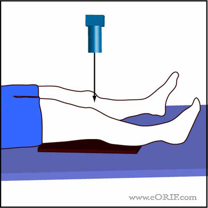 |
A/P Knee View
Position: supine with cassette under knee and femoral condyles parelll to cassette. Beam directed to point 1-2cm distal to the patella. |
||||||||
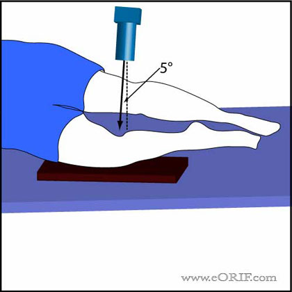 |
Lateral Knee View
Position: lateral with affected side down and flexed 30° at the knee. The contralateral leg is shifted posteriorly out of the way. Beam directed at knee joint with 5°cephalad angulation. |
||||||||
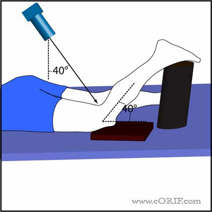 |
Tunnel View (Intercondylar notch view)
Position: prone, knee flexed 40° Beam directed caudally toward the knee joint at a 40° angle from vertical. |
||||||||
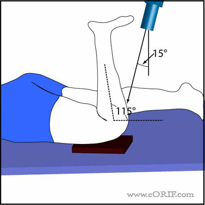 |
Sunrise View
Position: Prone; knee flexed 115° Beam directed toward patella with 15° cephalad angulation |
||||||||
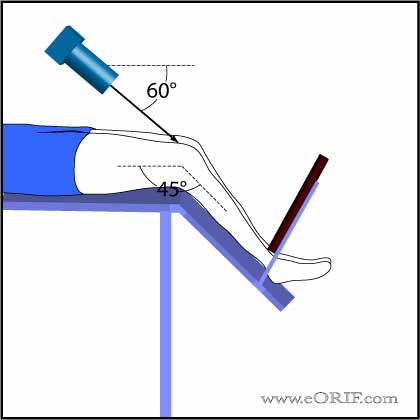 |
Merchant View
Position:supine knee flexed 45°; Beam directed caudally toward patella at a 60° angle (30° from the horixontal plane). (Merchant A, JBJS 1974; 56A:1391) |
||||||||
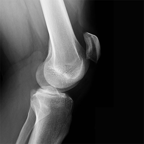 |
Patellar Alta, Patllar Baja
Image= lateral view of patient with patellar tendon rupture demonstrating patella alta. Insall-Salvati ratio
|
||||||||
| Radiology References |
